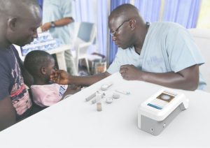
On December 18th, Noor van Driel successfully defended her MSc thesis titled “Automating malaria diagnosis: a machine learning approach: Erythrocyte segmentation and parasite identification in thin blood smear microscopy images using convolutional neural networks.”
In her thesis work, she proposes a two stage automated image classification strategy. Blood slides that were photographed at 20 X magnification were used in our experiments, allowing for a larger Field of View than regular thin film microscopy at 100 X. Erythrocytes are first localised and segmented by a Convolutional Neural Network, the architecture of which is based on U-Net, with some adaptations and improvements made for our purposes. The sensitivity and positive predictive value of the localisation were both 0.998, resulting in accurate cell counts. A transfer learning strategy, in which the existing VGG-16 network is used as a feature extractor and combined with a new fully connected layer to predict correct activations for our classification, is then used to classify the segmented erythrocytes as either infected with Plasmodium Falciparum parasite or healthy. Sensitivity and specificity of the predicted classification were 0.795 and 0.915 respectively. It is concluded that, although this method may not fully eliminate the need for trained experts, the algorithms proposed can be of great assistance in aiding the diagnostic decision making process.
The full thesis can be found here


